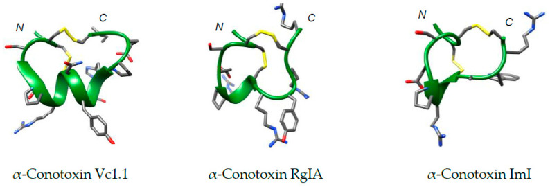
Figure 2.
Structures of α-conotoxins Vc1.1 (PDB: 2H28), Rg1A (PDB: 2JUT), and ImI (PDB: 1IMI) calculated from solution-state NMR data, provided from the Protein Data Bank (PDB) [34,43,45]. Structures were produced using Chimera [46]. These peptides share identical Loop I residues (GCCSDPRC) and possess variable Loop II primary sequences (full sequences are shown in Table 1). Peptide backbone shown in green, disulfide linkages in yellow and N- and C-termini are labelled.

