Figure 3.
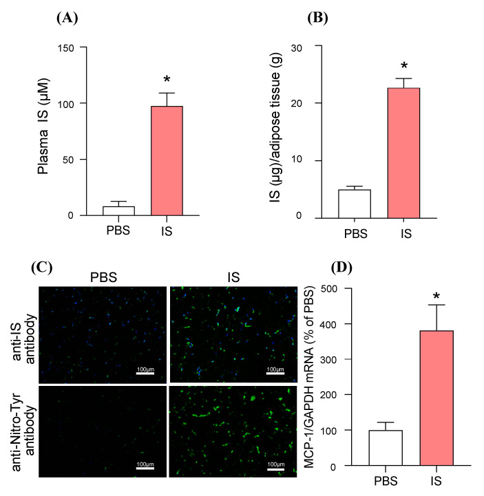
In vivo distribution of IS to adipose tissue and its MCP-1 inducing effect in IS-overloaded mice. The healthy mice were administrated with IS (100 mg/kg/day, ip). The control mice were administrated with the same volume of PBS. One hour after administration, the mice were anesthetized and blood, epididymal adipose tissue collected. IS levels in (A) plasma and (B) epididymal adipose tissue were measured by high-performance liquid chromatography (HPLC) methods. (C) IS accumulation (green) in epididymal adipose tissue was detected by immunofluorescence using anti-IS antibody. Immunofluorescent staining of nitrotyrosine (Nitro-Tyr: green) in epididymal adipose tissue was also shown. The section was also treated with DAPI (blue). Original magnifications: ×200. Scale bars represent 100 μm. (D) MCP-1 mRNA expression was determined by quantitative RT-PCR. Data are expressed as means ± SE (n = 4). * p < 0.05 compared with PBS-treated group.

