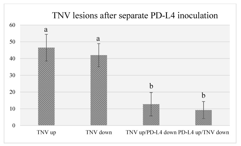
Figure 4.
Number of TNV lesions developed when the virus was inoculated alone on the adaxial (up) or abaxial (down) leaf surface, or when it was inoculated separately from PD-L4 in the opposite leaf surface. Different letters represent significant differences according to Fisher’s least significant difference test at p < 0.05. The error bars represent standard deviation.

