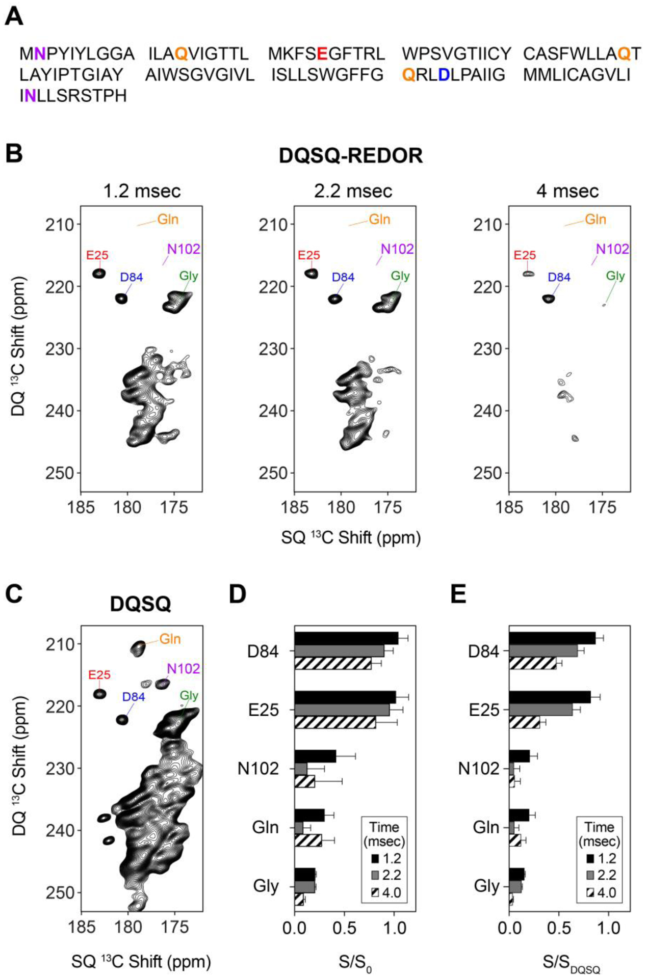
Figure 5.

(A) Primary sequence of EmrEE14Q with aspartate, asparagine, glutamate, and glutamine are color coded with residue types as in Figure 1. (B) DQSQ-REDOR and (C) DQSQ spectra of uniformly labeled 13C/15N EmrEE14Q in O-14:0-PC liposomes at a pH value of 5.0. The REDOR dephasing times in panel B are indicated on top of each spectrum. (D) Intensity ratios (S/S0) of select side chains (Asn, Asp, Gln, Glu) and backbone peaks (Gly) in the presence of 15N pulses (S) divided by those in the absence of 15N pulses (S0). (E) Intensity ratios (S/SDQSQ) of select side chains (Asn, Asp, Gln, Glu) and backbone peaks (Gly) in the presence of 15N REDOR pulses (S) divided by those in the DQSQ spectrum (SDQSQ).
