Figure 3.
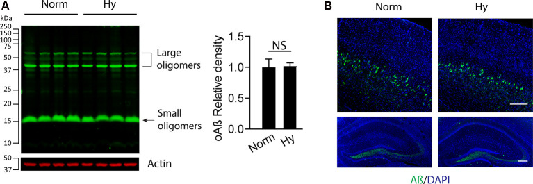
Antenatal hypoxia did not affect soluble Aβ accumulation in the brain cortex and hippocampus of 2-month-old 5xFAD offspring. The brain cortex was separated from the brain of 2-month-old 5xFAD offspring exposed to normoxia or hypoxia. (A) Western blotting was performed to detect the soluble Aβ levels with primary antibody against soluble Aβ (oAβ). Actin was used as internal control. Data are mean ± SEM. Student’s t test was applied to each data set. NS, not significant, n = 4. (B) Confocal imaging of soluble Aβ (green) distribution in the cortex (upper panel) and hippocampus (lower panel) in the brain of 2-month-old 5xFAD offspring exposed to normoxia or hypoxia. DAPI stains nuclei (blue). Scale bar: 200 μm.

