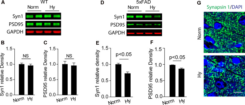
Figure 4.
Antenatal hypoxia resulted in synaptic loss in the brain cortex of 2-month-old 5xFAD offspring. (A–C) Western blotting analysis of the presynaptic marker Syn1 and postsynaptic marker PSD95 in the cortex of 2-month-old wild-type (WT) and (D–F) 5xFAD mice exposed to normoxia or hypoxia. Actin was used as internal control. Data are mean ± SEM. Student’s t-test was applied to each data set. n = 3–4. (G) Confocal images of brain slices from 2-month-old 5xFAD offspring exposed to normoxia or hypoxia stained with antibody against Synapsin 1 (green). DAPI stains nuclei (blue). Scale bar: 10 μm. NS, not significant.

