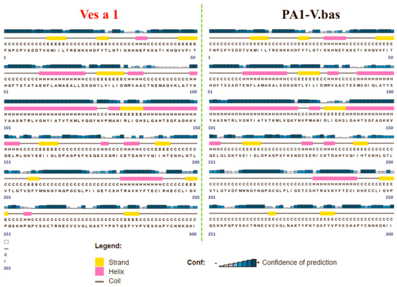
Figure 2.
Predicted secondary structures of Ves a 1 from V. affinis venom (Ves a 1) (red) and Ves a 1 from (V. basalis) venom (PA1-V.bas) (black), respectively: The H and pink rectangle represent a helical structure. The E and yellow rectangle represent a sheet structure. The C and black line represent a coil structure. The height of the blue bar represents the level of confidence in the predicted structure. The number indicates the amino acid order.

