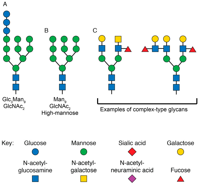
Figure 4.
Structures of glycans at various stages of N-linked glycosylation. (A) Graphical depiction of the glycan structure transferred to all N-linked glycosylation sites by the enzyme complex oligosaccharyltransferase in the rough ER. The three terminal glucose monomers are removed before exiting the ER. (B) Graphical representation of the high-mannose glycan at N-linked glycosylation sites as newly synthesized proteins enter the Golgi. For certain sites on HIV Env, this is the final form of the glycan. (C) Graphical representation of two complex-type glycans that are present on mature eukaryotic proteins. These are just two of the dozens of forms the final mature glycan structure can take. The sugar monomers are drawn according to the updated recommendations for symbol nomenclature for glycans [133].

