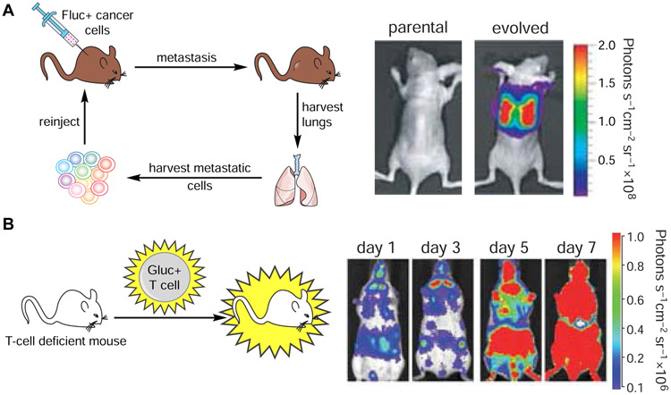
Figure 2. Noninvasive imaging in vivo with bioluminescent probes.
Luciferase-luciferin pairs have been routinely used to track cell migration and proliferation in vivo. (A) Imaging cancer cell proliferation and metastases. Mice were injected with Fluc-expressing MDA-MB-231 cells. Metastatic cells were harvested from lungs and re-injected into recipient mice. These cells exhibited a higher degree of metastatic outgrowth than the parental tumor. Figure reprinted with permission from Minn, et al., 2005 (B) Imaging T cell trafficking. Gluc-expressing T cells were injected into immunodeficient mice. T cell proliferation and homing were visualized over time via bioluminescence. Figure reprinted with permission from Santos et al., 2009.

