
Figure 1.
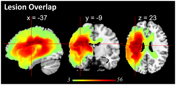
Lesion profile of the patient group. Subcortical regions and white matter, e.g. putamen, corpus callosum, thalamus, inferior occipito-frontal fasciculus, caudate, internal capsule, insular, had the largest probability of being damaged.
Official websites use .gov
A
.gov website belongs to an official
government organization in the United States.
Secure .gov websites use HTTPS
A lock (
) or https:// means you've safely
connected to the .gov website. Share sensitive
information only on official, secure websites.
Lesion profile of the patient group. Subcortical regions and white matter, e.g. putamen, corpus callosum, thalamus, inferior occipito-frontal fasciculus, caudate, internal capsule, insular, had the largest probability of being damaged.