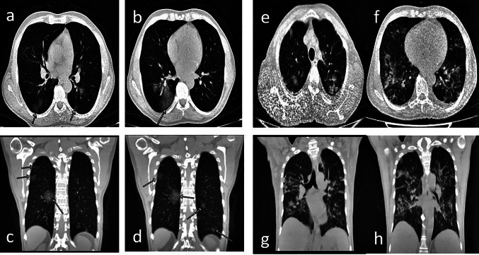
Fig. 1.
a–d Chest CT shows diffuse areas of diffuse ground-glass infiltrates in both lungs, scattered in a patchy style on March 15, 2020, after symptom onset. e–h Chest CT on March 31, 2020; In a comparative evaluation with CT of 15.03.2020, there is progression in pneumonic consolidations in both lungs. Interlobuler septal thickening in both lungs and pleural effusion increases reaching 10 mm on the left and 3 mm on the right in both hemithorax were observed as new findings

