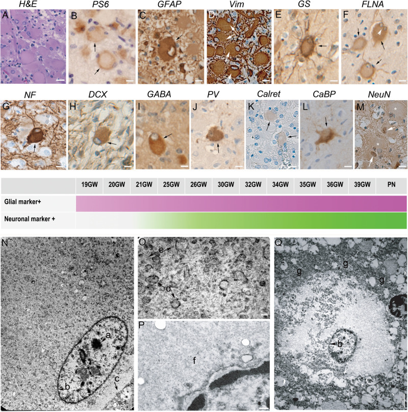
FIGURE 1.
Characterization of Giant cells. (A) Hematein-eosin staining of Giant cells displaying eosinophilic cytosols and flattened and decentered nuclei. (B) Immunostaining with antibodies to phosphorylated form or ribosomal PS6 (RPS6 R232H) of a couple of Giant cells. (C) Glial Fibrillary Acidic Protein immunopositive Giant cell; note the presence of GFAP negative cytomegalic neighboring cells. (D) Vimentin immunopositive Giant cell. some thin extensions were also immunopositive. (E) Glutamine synthetase antibodies stain the cytosol and the thin extensions of Giant cells. (F) Filamin A positive Giant cells, including a bi-nucleated one (arrow head). (G) Neurofilament 200 immunopositive balloon-like cell surrounded by immunonegative cytomegalic cells. (H–M) Giant cells immunopositive for different neuronal markers: DCX (Doublecortin), GABA (Gamma Amino Butyric Acid), PV (parvalbumin), Calret (Calretinin; only very slightly immunopositive as compared with neighbor cytomegalic cells), CaBP (Calbindin), NeuN (that stains mainly the nuclei). (N–Q) Electron microscopy pictures illustrating ultrastructure details of Giant cells displaying either neuronal (N, detail of the cytosol enlarged in O) or glial (Q, detail of the cytosol enlarged in P) features. a, nucleolus; b, nuclear membrane; c, cytoplasmic membrane; d, endoplasmic reticulum; e, filaments; f, smooth cytosol; g, accumulation of organelles at the periphery of the cytosol. Arrows in (A–M) point to immunopositive Giant cells; * in M indicate NeuN negative Giant cells. Figures were taken from 25GW (A,D,H,J,K,L), 26GW (B), 30GW (N–Q), 34GW (G,I,M) and 36GW (C,E,F); from cortex (E,G,I,N–Q), white matter nodule (A,C,D,J–K,M) and subependymal nodule (H). Scale bars: 10 μm (A–M), 1 μm (N), 100 nm (O,P), and 2 μm (Q). The frieze in the middle schematically represents the intensity of immunostaining of tuberous lesions throughout development. Arrows point to immunopositive Giant cells (B–M).

