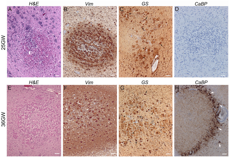
FIGURE 5.
Development of white matter nodules. (A–D) At 25GW hematein-eosin (A), vimentin (B), glutamine synthetase (C), and Calbindin (D) staining revealed the presence of Giant cells and dysmorphic astrocytes in white matter nodules. At this stage numerous SF cells distribute within the core of the nodule and at the periphery (as seen thanks to blue Nissl counterstaining), intermingled with cytomegalic cells. None cells were immunopositive for Calbindin (D). (E–H) At 36GW the nodule was less enriched in vimentin + (F) and GS + Giant cells (G), tend to be organized around a core of SF cells and displayed a sharp external “ring” of SF cells and dysmorphic neurons (white arrows) both calbindin + (H). Arrowheads and arrows in (C,G) point to some Giant cells and dysmorphic astrocytes respectively. Scale bars: 100 μm.

