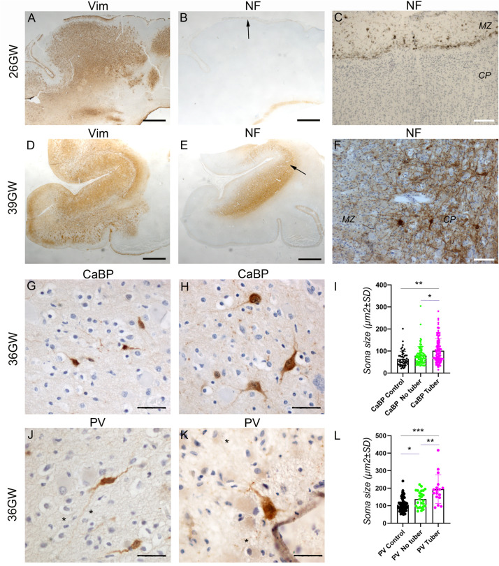
FIGURE 7.
Development of cortical TSC lesions: Neuronal features. (A–C) Pre-tuberal lesion on the cortex of a 26GW case, involving the presence of multiple vimentin positive Giant cells and dysmorphic astrocytes. However, NF200 immunopositive dysmorphic neurons are absent (B,C). Only the normal Brun layer of tangential fibers appears immunopositive in the marginal zone (C). (A,B) are close sections from the same cortex. The area depicted by arrow in (B) is enlarged in (C). (D–F) A typical tuber from a 39 GW case displaying an intense vimentin staining of Giant cells and dysmorphic astrocytes (D) and prominent NF200 immunostaining (E,F) of dysmorphic neurons and bundles of neurites, which accumulate mainly in the deep part of the tuber. (D,E) are close sections from the same cortex. The area depicted by arrow in (E) is enlarged in (F). (G–I) Calbindin immunostaining of cortex from a 36GW case. In non-tuberal cortex (G) the majority of immunopositive cells are of small size and display immature features. In tubers (H) frequent cytomegalic or dysmorphic calbindin positive cells were observed. (I) Quantification of cell soma size of stained neurons from 36 to 37GW control and TSC cases. Note that mean soma size is increased in tubers, with numerous cells displaying rater big soma size (n = 51 (control), 87 (no tuber) and 130 (tuber) cells). * and **p = 0.012 and 0.0004, respectively (Mann–Whitney test). (J–L) Parvalbumin immunostaining of cortex from a 36GW case. In non-tuberal cortex (J) the majority of immunopositive cells are of small size and display immature features. In tubers (K) a few cytomegalic or dysmorphic PV positive cells were observed (* tags Giant cells immunonegative). (L) Quantification of cell soma size of stained neurons from 36 to 37GW control and TSC cases. Note that mean size is increased in tubers, with some cells displaying rather big soma size (n = 63 (control), 26 (no tuber) and 17 (tuber) cells). *, **, and *** p = 0.036, 0.0013, and 0.0001 respectively (t-test). SG, subpial granular layer, MZ, marginal zone, CP, cortical plate. Scale bars: 2 mm (A,B,D,E), 100 μm (C,F), and 50 μm (G,H,J,K).

