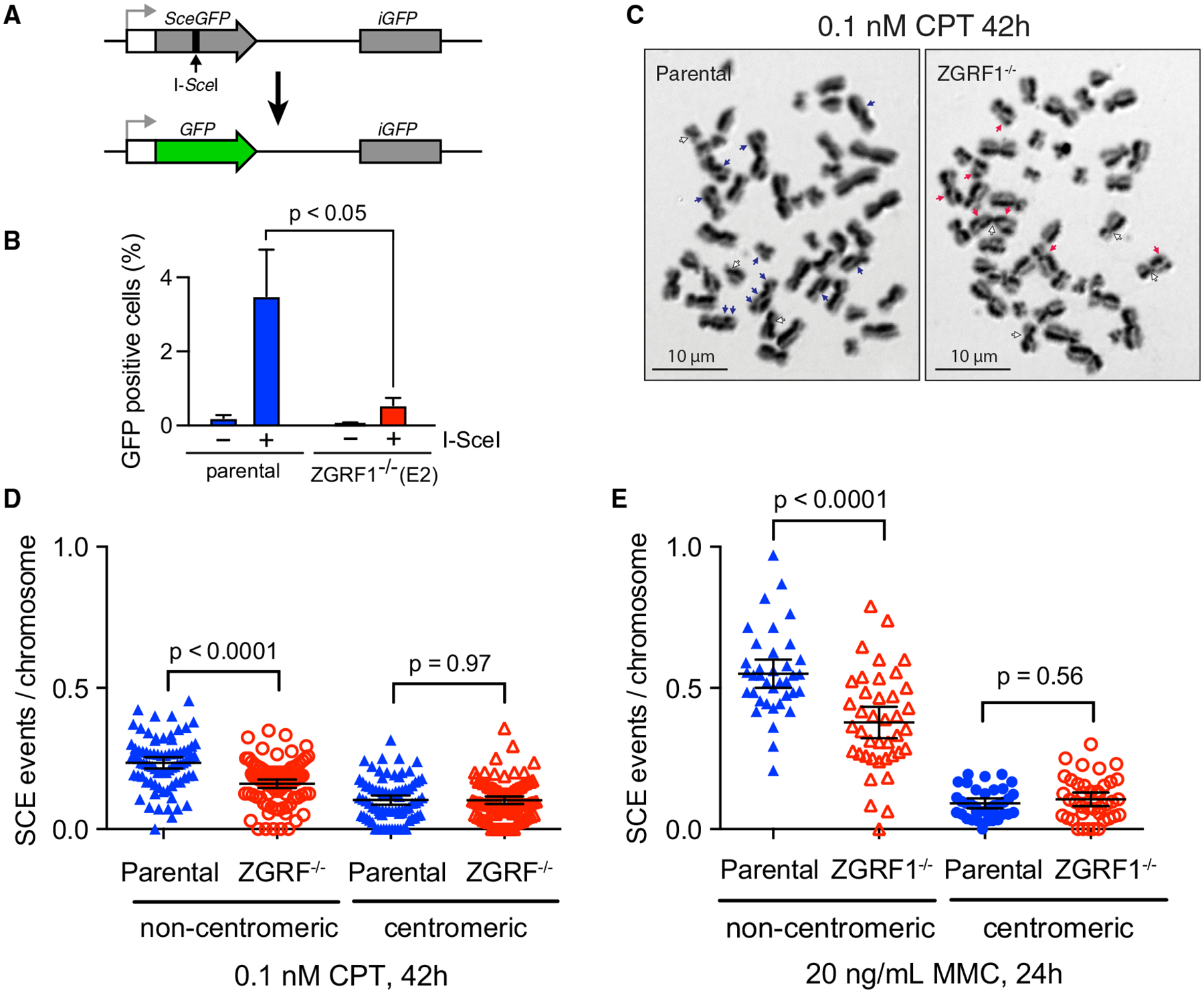
Figure 4. ZGRF1 Contributes to Homologous Recombination.

(A) Schematic illustration of the DR-GFP assay. A recognition site of the I-SceI meganuclease has been integrated into the open reading frame (ORF) of the GFP gene (SceGFP), thereby disrupting the ORF, and a truncated GFP gene (iGFP) fragment with the correct ORF sequence is placed downstream in the construct. Repair of the cleaved I-SceI site by gene conversion using the downstream iGFP as a template results in a functional GFP gene that is measured using flow cytometry. White box, promoter; gray arrow, transcription start site.
(B) ZGRF1 promotes gene conversion. The percentage of GFP-positive cells with (+) or without (−) I-SceI expression is shown for U2OS DR-GFP parental and ZGRF1−/− cells (p < 0.05, multiple t test). Error bars indicate SEM (n = 4).
(C) Representative images of sister chromatid exchange events (SCEs) in metaphase spreads in cells treated with 0.1 nM CPT for 42 h. Blue and red arrows mark non-centromeric exchanges for HCT116 parental and ZGRF1−/− cells, respectively, while white arrows mark centromeric exchanges.
(D and E) ZGRF1−/− cells show a decrease in the number of non-centromeric SCEs per chromosome compared with HCT116 parental cells. Quantifications of SCE frequencies in HCT116 parental and ZGRF1−/− cells treated with 0.1 nM CPT for 42 h or 20 ng/mL MMC for 24 h are shown. Two independent experiments were performed. N ≥ 35 metaphase spreads and 674 chromosomes per condition.
Error bars show 95% confidence intervals. p values were calculated using Mann-Whitney U tests.
