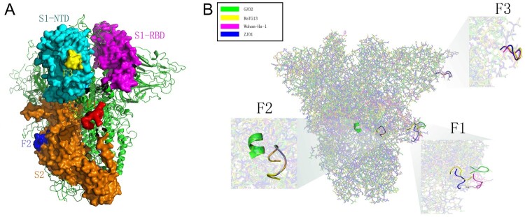
Figure 5.
Spatial location and protein structure of potential FCS. (A) The spatial position of F1-3 on S protein. F1 is located at S1/S2, F2 at S2 and F3 at the NTD of S1. (B) Differences in the tertiary structure of the protein at the F1-3 sites of GZ02, RaTG13, Wuhan-Hu-1 and ZJ01. The difference between ZJ01 and Wuhan-Hu-1 may be caused by the mutation of ZJ01 near the FCS.

