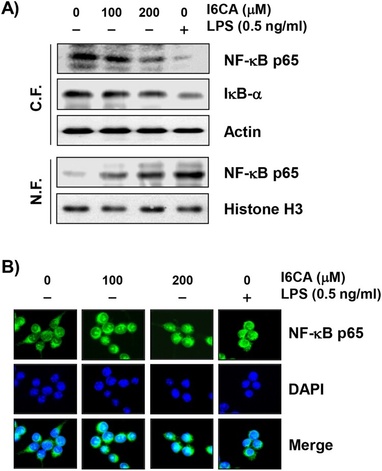
Figure 7.
Effect of I6CA on the expression of NF-κB, and IκB-α. RAW 264.7 cells were treated with I6CA or LPS for 24 h. (A) Cytoplasmic and nuclear proteins were isolated for analysis of NF-κB, IκB-α, and p-IκB-α expression, and Western blot analysis was performed. Analysis of actin and histone H3 expression was performed to confirm the protein loading of each fraction extract. (B) The localization of NF-κB/p65 (green) was visualized by fluorescence microscopy. These cells were also stained with DAPI to visualize the nuclei (blue).

