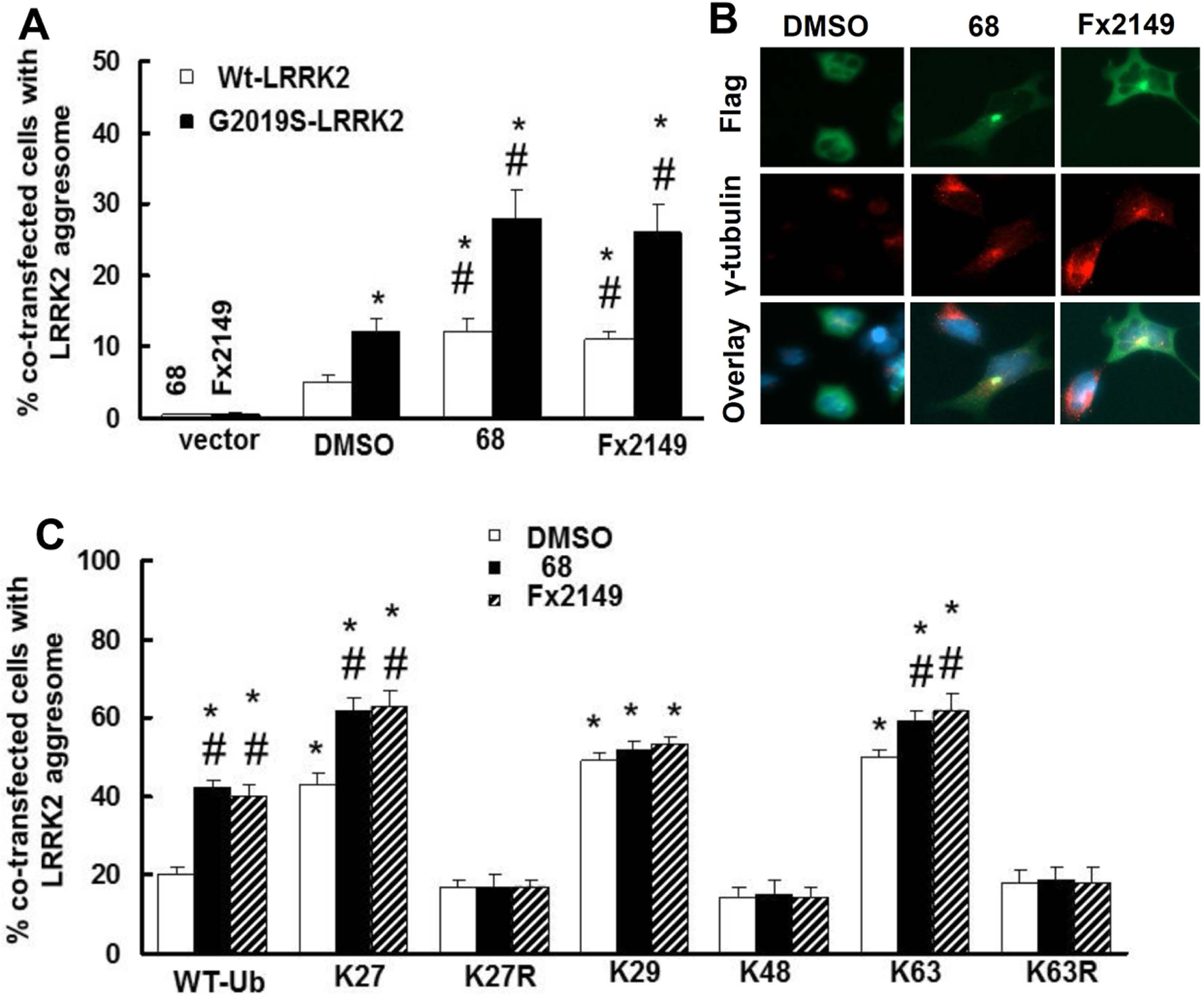
Fig. 3. 68 increased G2019S-LRRK2 ubiquitination predominantly via K27-linkages.

HEK293T cells were co-transfected with Flag-G2019S-LRRK2 and various HA-tagged ubiquitin constructs for 48 h, then treated with 68 (100 nM) for 24 h. Cell lysates were subjected to ubiquitination assays. A, Representative blots from three repeated ubiquitination assays. B, quantification of density of ubiquitination by NIH image J software. P<0.05 by ANOVA, * vs cells expressing WT-LRRK2 treated with 0.2% DMSO; # vs vehicle control of each genotype of cells.
