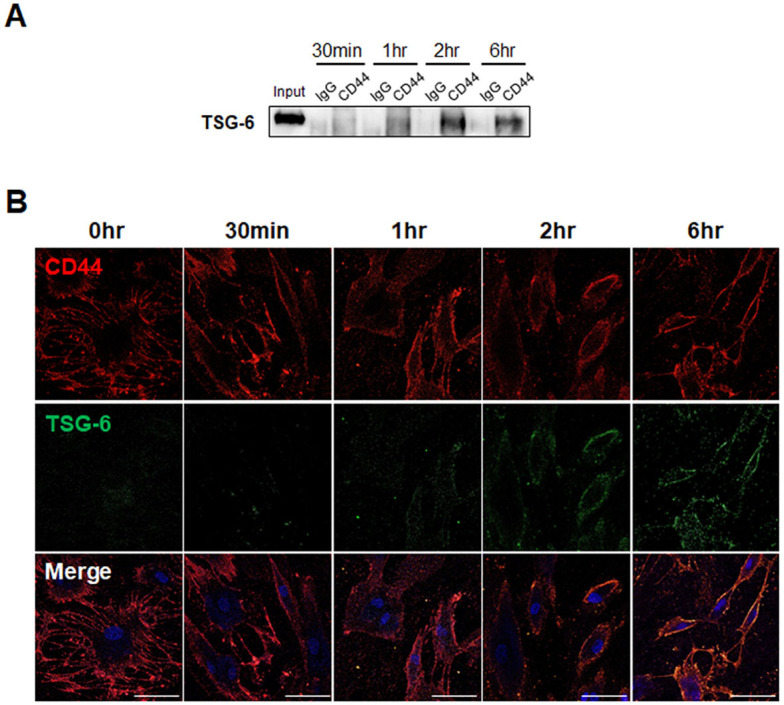
Fig. 1.
Interaction of TSG-6 with CD44 receptor in human pHSCs. (A) Immunoblots with TSG-6 in the immunoprecipitated pHSC lysates with CD44. pHSCs were treated with TSG-6 for 30 min, 1 hour, 2 hours, and 6 hours, respectively and then immunoprecipi-tated with CD44 antibody or IgG as negative control. Input sam-ples containing the same amount of TSG-6 protein used for in vitro treatment were used as positive controls. Data shown represent one of three experiments with similar results. (B) Immunofluo-rescence staining for FITC-conjugated TSG-6 (green) and CD44 (red) in pHSCs treated with TSG-6. DAPI was used in staining for nucleus. Representative images obtained from three experiments with similar results are shown (scale bar = 50 µm).

