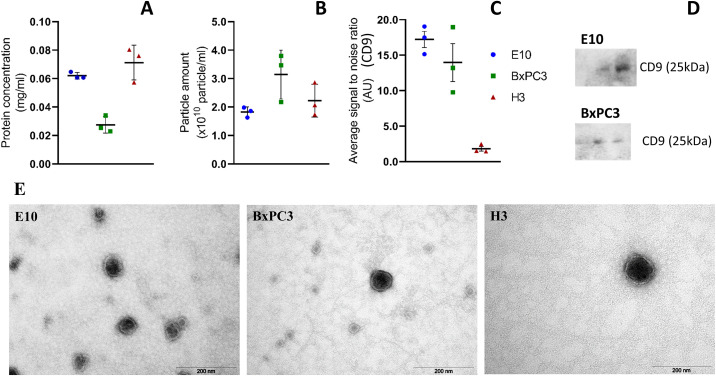
Fig 1. Characterization of the small EVs isolated from the cell culture supernatant of the E10, BxPC3, and H3 cell lines.
EVs were studied by protein concentration measurements (A), NTA for particle quantification (B), immunoaffinity capture targeting CD9 (C), and Western blot for the detection of CD9 in three sequential SEC fractions (20 μl per well) (D). Samples were negatively stained with 4% uranyl acetate (aqueous) for analysis by TEM (E). Data republished from Guerreiro et al [23].

