Fig. 4.
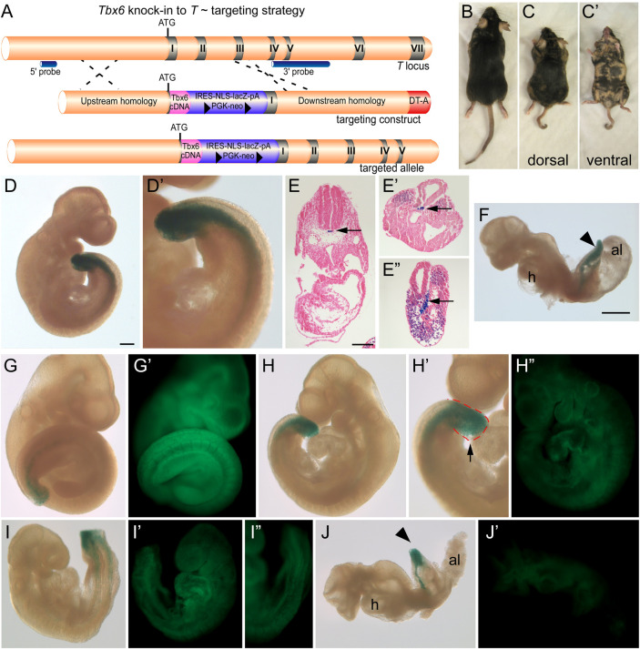
Tbx6 knockin T targeting strategy and chimera phenotypes. (A) Schematic of the targeting strategy to knock the Tbx6 cDNA into the T locus at the initiating methionine TTbx6ki. The positions of the IRES-lacZ-PGK-neo positive selection cassette, the diphtheria toxin A (DT-A) negative selection cassette, and 5′ and 3′ external probes for genotyping are indicated. (B,C) Dorsal views of littermate chimeric mice derived from injecting TTbx6ki/+ ES cells into C57 blastocysts. Mice in panels B and C represent ∼10% and 40% chimerism, respectively. (C′) Ventral view of chimera shown in panel C. Note the short axis and kinky tail of the higher percentage chimera (panel C) compared to its littermate (panel B). (D,D′) β-galactosidase staining of a chimeric embryo and representative sections (E–E″) showing the presence of the lacZ reporter activity in the PS and notochord (arrow), indicative of the T expression domains. (G–J) Chimeric embryos resulting from TTbx6ki/+ ES cell injections into GFP-expressing blastocysts were stained for β-galactosidase activity and imaged in bright field and GFP fluorescence (G′–J′). Panels G to J represent low (panel G,G′) to high percentage (panel J,J′) contribution of the TTbx6ki/+ ES cells in chimeric embryos as shown by reduced GFP from panels G′ to J′. Developmental defects include abnormal tail and somite morphology (H,I) and shortened axis and failure to turn (J) in the higher percentage chimeras. Magnification bars represent 200 μm (D,G–I), 150 μm (E), 700 μm (F,J).

