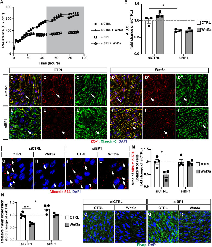
Fig. 6.
Fgfbp1 knockdown impairs Wnt-induced paracellular and transcellular barrier properties in brain endothelial cells in vitro. (A) TEER of a monolayer of mBECs transfected with either an siRNA targeting Fgfbp1 (siBP1) or a scramble siRNA (siCTRL). The gray area represents the period of time during which cells were treated with either recombinant Wnt3a or vehicle (control). (B) Quantification of the area under the curve (A.U.C.) during Wnt3a treatment (gray area in A). Each dot represents an independent experiment. siBP1-transfected cells have reduced TEER compared with siCTRL-transfected cells. Data are mean±s.e.m. *P<0.05; one-way ANOVA. (C-F″) Immunofluorescence for claudin 5 (green) and ZO-1 (red) in mBECs transfected with either siBP1 or siCTRL either with or without Wnt3a. There is no difference in claudin 5 localization, but ZO-1 expression is reduced in siBP1-transfected compared with siCTRL-transfected mBECs. (G-L) Immunofluorescence for the uptake of Albumin-Alexa 594 (red dots, white arrows) in siCTRL or siBP1-transfected mBECs with or without Wnt3a. DAPI (blue) labels nuclei. (M) Quantification of Albumin-Alexa 594 uptake by siCTRL- or siBP1-transfected mBECs with or without Wnt3a. Each dot represents an independent experiment. Wnt3a reduces the uptake of Albumin-Alexa594 in siCTRL, but not in siBP1-transfected mBECs. Data are mean± s.e.m. *P<0.05; one-way ANOVA. (N) Fold change expression of Plvap mRNA in siCTRL or siBP1-transfected mBECs either untreated or treated with Wnt3a. Each dot represents an independent experiment. Plvap expression is reduced by Wnt3a treatment in siCTRL-transfected but not siBP1-transfected mBECs. Data are mean±s.e.m. *P<0.05, **P<0.005; one-way ANOVA. (O-R) Immunofluorescence for Plvap (green) and DAPI (blue) in siCTRL or siBP1-transfected mBECs with or without Wnt3a. Scale bars: 20 µm.

