Fig. 8.
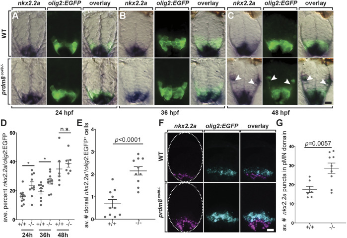
pMN cells prematurely express nkx2.2a in prdm8 mutant embryos. (A-C) Representative transverse sections of trunk spinal cord (dorsal up) showing prdm8 RNA (blue) and olig2:EGFP (green) expression. Developmental stages are noted at the bottom. Arrowheads indicate dorsally migrated oligodendrocyte lineage cells. (D) The area of nkx2.2a expression in the pMN domain is greater at 24 (n=10) and 36 hpf (co49, n=9; wild type, n=10) in prdm8 mutants embryos compared with controls and there is no difference at 48 hpf (co49 n=6; wild type, n=7). (E) prdm8co49−/− (n=10) have more dorsal OPCs (nkx2.2a+/olig2:EGFP+) than wild-type embryos (n=10) at 48 hpf. (F) Representative transverse trunk spinal cord sections obtained from 28 hpf embryos processed for fluorescent ISH to detect olig2 (blue) and nkx2.2a (pink) mRNA. Dashed ovals outline the spinal cord. (G) More nkx2.2a puncta are located within the olig2+ pMN domain of prdm8co49−/− embryos (n=9) compared with wild-type embryos (n=7). Data are mean±s.e.m. with individual data points indicated. Statistical significance was evaluated by an unpaired, two-tailed Student's t-test. n.s., not significant. *P<0.05. Scale bars: 10 μm.

