Fig. 3.
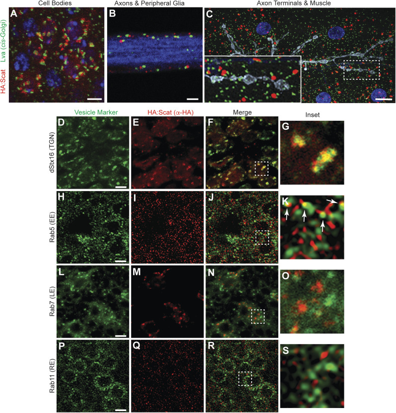
Scat localizes to the TGN in MN cell bodies. (A–C) Scat localizes to a structure adjacent to the cis-Golgi in (A) motor neuron cell bodies, (B) peripheral glia, and (C) body wall muscle. (A,B) Ventral ganglia and (C) body wall muscle preps from wandering third instar larvae expressing inducible HA:scat under control of the tubulin-Gal4 driver were stained with antibodies targeting the HA tag (red) and the cis-Golgi marker, Lva (green). Single focal planes are shown in A and B while C is a maximum Z-projection. HA:Scat localizes to the motor neuron cell body but not peripheral axons or axon terminals. Most HA-positive structures are adjacent to the Lva-positive cis-Golgi. Blue is DAPI (DNA) in A and C and Hrp (axon) in B. Grey in C is Hrp (axon). Scale bars are 2.5 µm in A and 10 µm in B and C. (D–S) tub-Gal4>HA:scat animals were counterstained with antibodies targeting the HA tag (red) and the indicated marker (green). Images shown are single focal planes through MN cell bodies in the larval ventral ganglion. Vesicle trafficking markers shown are the TGN marker, Syntaxin 16 (D–G), the early endosome marker, Rab5 (H–K), the late endosome marker, Rab7 (L–O), and the recycling endosome marker, Rab11 (P–S). Larvae showing the colocalization of HA:Scat with Rab5 and Rab11 were fixed with Bouin's reagent, which provided much better signal to noise. Larvae showing HA:Scat with dStx16 and Rab7 were fixed with paraformaldehyde. The boxed areas indicated in the merged images (F, J, N and R) are shown in G, K, O and S (respectively). The arrows shown in K are indicating localization of HA:Scat in spots immediately adjacent to Rab5. Scale bars in D, H, L and P are 2.5 µm.

