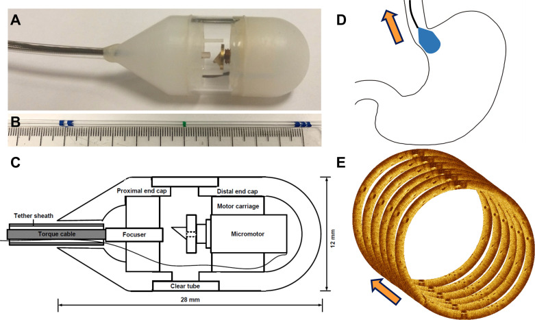
Figure 1.
(A) Photograph of tethered optical coherence tomography (OCT) capsule constructed using lubricious material with a 30° proximal taper for ease of retrieval. (B) Tether markings every 5 cm indicating distance from incisors. (C) Schematic showing micromotor rotary optical scanner and other components. (D) Cartoon showing capsule travelling from gastric cardia into distal oesophagus during a pullback image acquisition. (E) Illustration showing multiple cross-sectional images acquired in rapid succession during capsule pullback to obtain volumetric data for subsurface en face and cross-sectional visualisation.

