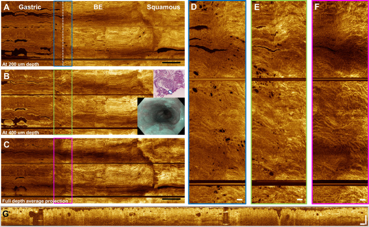
Figure 4.
Tethered capsule optical coherence tomography (OCT) from a patient with C2M4 non-dysplastic Barrett’s oesophagus (BE). Inset shows narrow band imaging view from a previous endoscopy. (A) En face OCT at 200 µm depth, (B) 400 µm depth and (C) full depth projection. Scale bars 1 cm. Some longitudinal pullback non-uniformity can be observed in the BE segment. (D–F) Enlargements showing glands and mucosal pattern at the gastro-oesophageal junction (GEJ). Scale bars 1 mm. (G) Cross-sectional OCT from the GEJ showing atypical glands. Scale bar 500 µm. Biopsy at the GEJ (inset) from a prior oesophagogastroduodenoscopy (9 months earlier) shows a large dilated cardiac gland (arrow) with smaller peripheral glands from the superficial mucosa.

