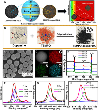
Fig. 1. Synthesis and characterization of TEMPO-doped PDA.

(A) Schematic illustration of the TEMPO-doped PDA with narrower bandgap and improved light absorption ability compared to conventional PDA. (B) Polymerization of dopamine and TEMPO, together with their molecular structures and powder photographs. (C) SEM image of PDA-3. (D) EELS mapping analysis of PDA-3 (Scale bars, 100 nm). (E) XPS survey spectra of PDA-i (i = 0 to 3). a.u., arbitrary units. (F) C 1s peaks, (G) N 1s peaks, and (H) O 1s peaks in XPS spectra of PDA-3.
