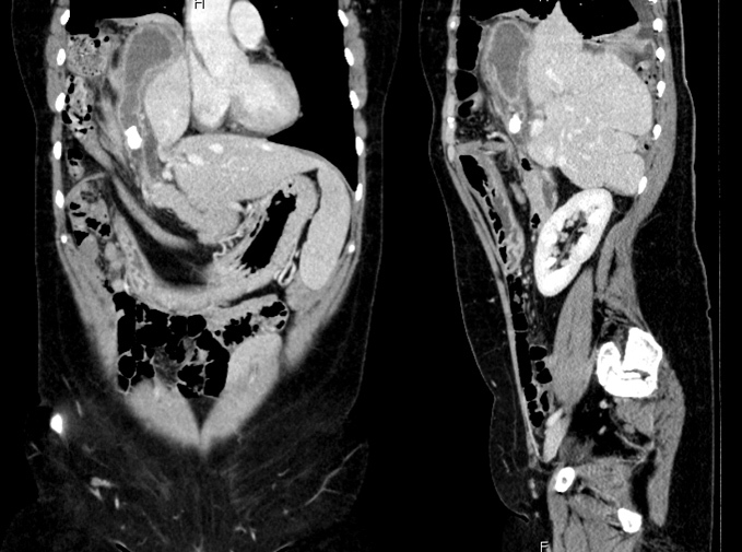
Figure 1.

Computed tomography showing a large right-sided diaphragmatic hernia, with intrathoracic herniation of the right hepatic lobe and gallbladder which is distended, with thickened walls and a large gallstone (18 mm) in the infundibular region. Another gallstone (5 mm) can be identified in the common bile duct
