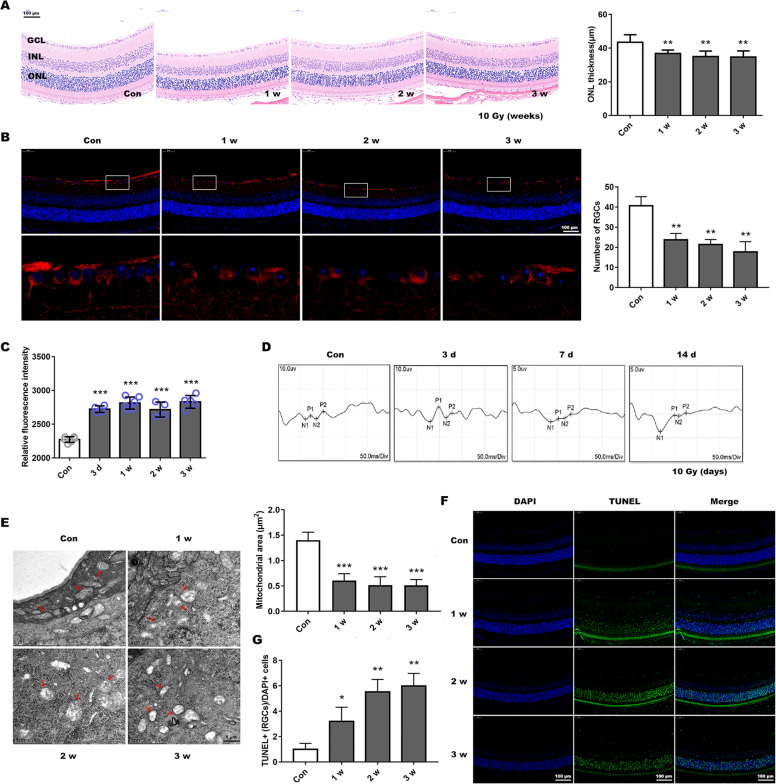
Fig. 6. X-ray-induced mitochondrial dynamic changes and visual function damage in BALB/c mice.
a Hematoxylin and eosin staining showed retinal structures changes after 10 Gy exposure and analyzed the outer nuclear layer (ONL) thickness. GCL ganglion cell layer, INL inner nuclear layer. Scale bar is 100 µm. b RGCs and neuronal axons identified using Tuj1 antibodies by immunofluorescence with retinal paraffin sections. Lower panel presents a fractionated gain of the image in the white rectangle of the upper panel. The number of RGCs at different post-radiation time (1, 2, and 3 weeks) were then calculated. Bar, 100 µm. c Retinal ROS generation was determined by DCFH-DA fluorescence intensity quantification after radiation for 3 d or 1–3 weeks. d Visual function was evaluated by VEP recording after 3, 7, or 14 d irradiation. e Mitochondrial micromorphology was determined by transmission electron microscopy. The red arrows indicate markedly morphologic mitochondria, and mitochondrial area was measured. Bar, 1 µm. f Retinal paraffin sections were stained by TUNEL to observe cellular apoptosis, and g TUNEL-positive RGCs were counted. Bar is 100 µm. Data are presented as the mean ± SD (n = 3–6), *P < 0.05; **P < 0.01; ***P < 0.001.

