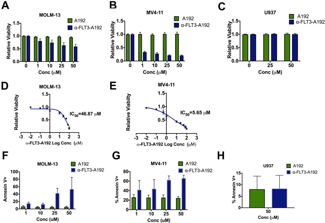
Figure 4: α-FLT3-A192 has anti-leukemic activity in AML cells.
A-C) A trypan blue viability assay was performed in MOLM-13, MV4–11 (FLT3 ITD+) and U937 (FLT3 negative) cells treated with α-FLT3-A192 or an A192 control for 72 hours. The number of live cells was normalized to untreated cells. Data represented as mean ± SD, n=6. D-E) An IC50 of α-FLT3-A192 was measured using alamar blue staining in MOLM-13 and MV4–11 cells at 72 hours post treatment with the increasing concentration of α-FLT3-A192 and plotted based on non-linear regression. F-H) Apoptosis was measured by flow cytometry in MOLM-13, MV4–11 (FLT3 ITD+) and U937 (FLT3 negative) cells at 72 hours post treatment with α-FLT3-A192 or control A192 using APC conjugated Annexin V stain and normalized to untreated cells. Data represented as mean ± SD, n=3.

