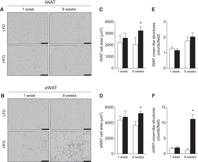
Fig. 2.
Effects of 1 and 8 wk of HFD on adipocyte size and white adipose tissue leukocyte infiltration. Representative images of hematoxylin-and-eosin (H&E) stained iWAT (top) and eWAT (bottom) from LFD- and HFD-fed mice imaged at ×40 magnification (A and B), cross-sectional adipocyte area (C and D), crown-like structure quantification (E and F) (n = 4–6/experiment). Open bars denote LFD-fed mice, while solid bars denote HFD-fed mice. Scale bars are 100 μm. Data are expressed as means ± SE *P < 0.05 compared with LFD group. eWAT, epididymal white adipose tissue; iWAT, inguinal white adipose.

