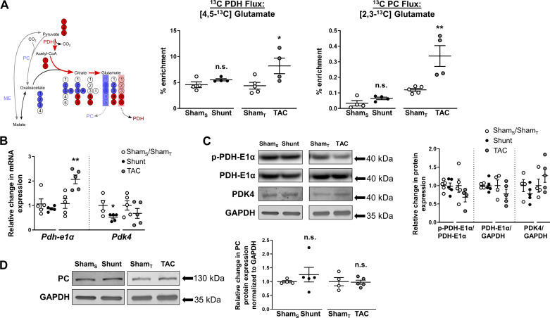
Fig. 6.
Effects of Shunt and transverse aortic constriction (TAC) on cardiac tricarboxylic acid (TCA) cycle activity. A, left: following [U-13C]glucose administration, [1,2,3-13C]pyruvate arises from glycolysis, which can enter the TCA cycle via pyruvate dehydrogenase (PDH) or through anaplerotic reactions via pyruvate carboxylase (PC) or malic enzyme (ME). [4,5-13C]glutamate was used as a readout for PDH activity and [2,3-13C]glutamate for anaplerosis. Red circles, 13C-labeled carbons arising from PDH; blue circles, from anaplerotic reactions. A, right: 13C enrichment of [4,5-13C]glutamate and [2,3-13C]glutamate in Shunt, TAC, or respective control hearts. ShamS, control group for Shunt; ShamT, control group for TAC. B: cardiac mRNA expression of pyruvate dehydrogenase-E1α (Pdh-E1α) and pyruvate dehydrogenase kinase-4 (Pdk-4). C: cardiac protein expression of phospho-PDH-E1α (S293, p-PDH-E1α), total PDH-E1α, and PDK-4. D: protein levels of PC. GAPDH served as internal control. n = 4–5 Mice/group. Data are presented as means ± SE. *P < 0.05 and **P < 0.01 between Shunt/TAC and the respective Sham controls. ns, Not significant by unpaired Student’s t test. Gaps in the x-axis indicate that Shunt and TAC experiments were performed and analyzed separately.

