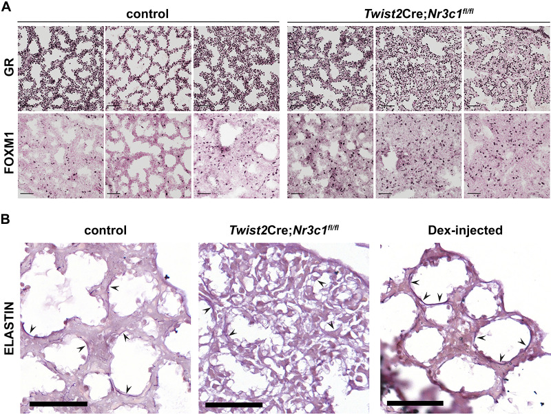
Fig. 9.
Dexamethasone increased elastin staining in peripheral saccules of embryonic lung A: embryonic day (E)18.5 lung tissue from control or Twist2Cre;Nr3c1fl/fl mice were stained with glucocorticoid receptor (GR) and FOXM1 antibodies. Each column represents lung tissue from an individual animal. Note decreased GR staining in mesenchyme of Twist2Cre;Nr3c1fl/fl mice and increased FOXM1+ cells compared with controls. Scale bars = 50 µm. B: lung tissues were obtained at E18.5 and stained for elastin. Wild-type mouse dams were injected with saline (left) or 1 mg/mL dexamethasone (Dex; right) in vivo 24 h before euthanize at E18.5. Middle: elastin staining of lung tissue following mesenchymal GR deletion (Twist2Cre;Nr3c1fl/fl). Arrowheads denote elastin staining in developing saccules of the distal lung. Note decreased elastin staining in Twist2Cre;Nr3c1fl/fl lungs and increased staining in the wild-type lung treated with antenatal Dex. Images are representative of n = 3 control lungs; n = 3 Twist2Cre;Nr3c1fl/fl lungs; and n = 7 Dex-injected wild-type lungs. Scale bar = 100 μm.

