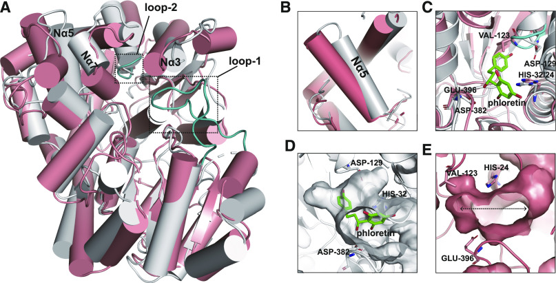
Figure 7.
Comparison of the Structures of UGT708C1-Phloretin and CGT TcCGT1.
(A) Overall structural comparison of UGT708C1-phloretin (gray cartoon) and TcCGT1 (salmon cartoon). Loop-1 and loop-2 in the structure of TcCGT1 are shown in cyan and indicated by dotted boxes. The positions of Nα3, Nα5, and Nα7 in the structure of UGT708C1 are labeled.
(B) The helix of TcCGT1 (salmon cartoon) corresponding to the Nα5 of UGT708C1 (gray cartoon) is significantly shifted toward the outside of the binding pocket.
(C) Residue Val123 on loop-2 in the structure of TcCGT1 is located in the position of phloretin in the structural model of UGT708C1-phloretin. Phloretin is shown as green sticks, and all atoms are colored according to element (carbon, green; oxygen, red).
(D) The UGT708C1 acceptor binding pocket is shaped like a curved “L.” Phloretin binds to the pocket in a bent state. Phloretin is shown as green sticks, and all atoms are colored according to element (carbon, green; oxygen, red).
(E) The TcCGT1 acceptor binding pocket is shaped like a horizontal “I.” The substrate binds to the spacious pocket.

