
Figure 1.
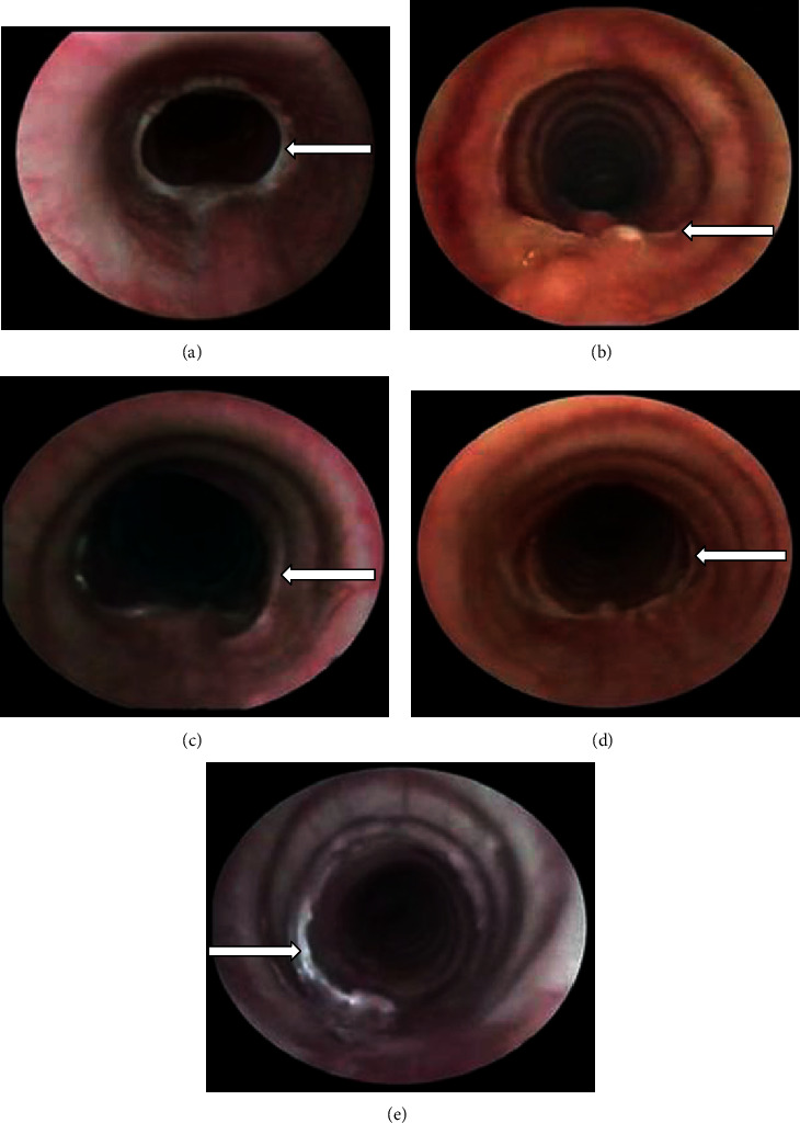
Representative endoscopic view of one animal in each group showing the scar formed in the tracheal anastomosis 4 weeks after treatment. (a) Group I (control): tracheal scar with a concentric membrane of fibrous tissue, which caused grade II stenosis (arrow). (b) Group II (hyaluronic acid), (c) group III (collagen-PVP), and (d) group IV (mixture of hyaluronic acid+collagen-PVP) showing anastomosis scars (arrow) without inflammation or oedema and with 100% patency. (e) Group V (mitomycin C): mild granulation tissue over the mucosa of the tracheal anastomosis (arrow) and 100% of the tracheal lumen.