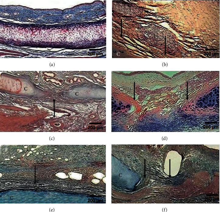
Figure 2.

Microphotographs of tracheal scars (Masson's trichrome staining, 2x magnification). (a) Normal histology of the trachea. (b) Group I (control): severe inflammation (I) and disorganized collagen fibres (arrow) around the cartilage (C). (c) Group II (hyaluronic acid), (d) group III (collagen-PVP), (e) group IV (mixture of hyaluronic acid+collagen-PVP), and (f) group V (mitomycin C): well-organized collagen fibres (arrow), mild-to-moderate inflammatory infiltration, and neoformation vessels.
