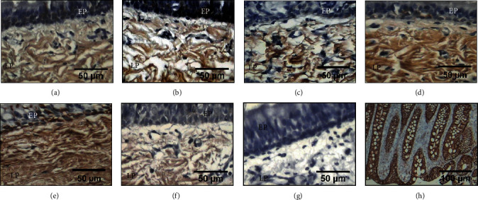
Figure 4.

Immunohistochemical detection of decorin in the epithelium (EP) and lamina propria (LP) from tracheal scars presurgery (a) and after treatment with WHMs: (b) group I (control), (c) group II (hyaluronic acid), and (f) group V (mitomycin C) showing light brown immunostaining. (d) Group III (collagen-PVP) and (e) group IV (mixture of hyaluronic acid+collagen-PVP) showing strong brown immunostaining. (g) Tracheal tissue as a negative control. (h) The human colon was the positive control.
