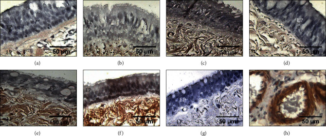
Figure 5.

Immunohistochemical detection of MMP1 in the epithelium (EP) and lamina propria (LP) from tracheal scars posttreatment with WHMs. Immunostaining in light brown showing the expression of MMP1 in (a) presurgery samples and in (c) group II (hyaluronic acid), (d) group III (collagen-PVP), and (e) group IV (mixture of hyaluronic acid+collagen-PVP). Strong brown immunostaining indicates MMP1 expression in (b) group I (control) and (f) group V (mitomycin C). (g) Tracheal tissue as a negative control. (h) The vascular endothelium of rat lung tissue was the positive control.
