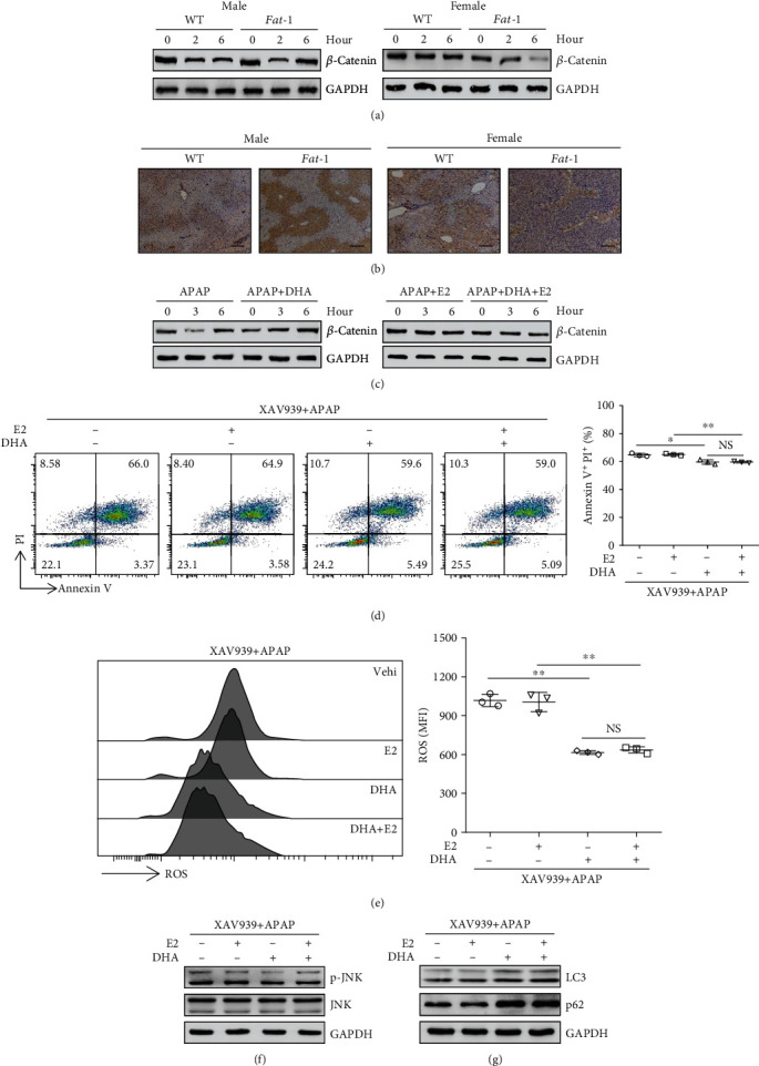
Figure 4.

β-Catenin signaling is involved in the differential effect of n-3 PUFAs on APAP hepatotoxicity between male and female mice. (a, b) APAP (400 mg/kg) was intraperitoneally injected to male or female WT and fat-1 mice (n = 5). Subsequently, the liver tissues were collected at 6 hours post-APAP injection. (a) The protein level of β-catenin in the liver tissues was evaluated by western blotting. (b) Immunohistochemical staining for β-catenin was determined in the liver tissue. Scale bars = 100 μm. (c) HepaRG cells were pretreated with 50 μM DHA without 100 nM E2 before stimulation with 20 mM APAP. Western blot assay of β-catenin was performed at the indicated time point after APAP stimulation. (d–g) HepaRG cells were pretreated with 2 μM XAV939 for 2 hours combined with 100 nM E2 in the presence or absence of 50 μM DHA. Subsequently, the cells were stimulated with APAP for another 24 hours. (d) Apoptosis was measured by Annexin V-PI staining, followed by FACS analysis. (e) The ROS level in the cells was analyzed by flow cytometry labeling with fluorescent probe DCFH-DA at 6 hours following APAP administration. (f) Phosphorylation of JNK expression in the APAP-treated cells was evaluated at 6 hours following APAP administration by immunoblotting analysis. (g) The expression of LC3 and p62 was determined by immunoblotting analysis. ∗p < 0.05, ∗∗p < 0.01. NS: not significant. The data represent three independent experiments with similar results.
