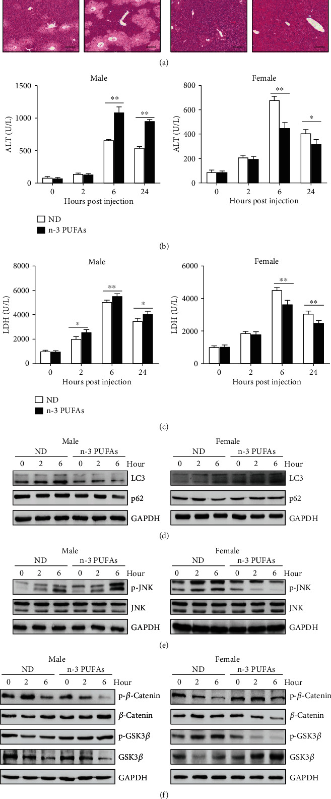
Figure 6.

Sex-specific effect of exogenous n-3 PUFAs on APAP-induced liver damage. APAP (400 mg/kg) was intraperitoneally injected into male or female WT mice fed with normal diet or n-3 PUFA-enriched diet (n = 5). (a) 24 hours after APAP injection, histological analysis of mouse livers was performed by H&E staining. Scale bars = 100 μm. (b, c) Serum ALT and LDH levels at different time points post-APAP injection were measured. (d) The protein levels of LC3 and p62 in liver tissues were determined by immunoblotting analysis. (e) Phosphorylation of JNK expression in livers was evaluated by immunoblotting analysis. (f) The phosphorylation of β-catenin and GSK3β was determined by immunoblotting analysis at the indicated time point after APAP administration. ∗∗p < 0.01. The data represent three independent experiments with similar results.
