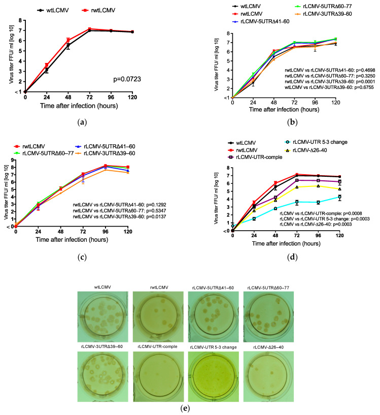
Figure 3.
Viral growth properties in Vero and A549 cells: Confluent monolayers of Vero and A549 cells were infected with wtLCMV or rLCMVs at a multiplicity of infection (MOI) of 0.001 per cell. Cells were washed three times with medium after a one-hour adsorption period, and 1 mL medium was added to each well. Supernatant samples were collected at 0, 24, 48, 72, 96, and 120 h postinfection. The supernatants were centrifuged at 8000× g for 5 min to remove cell debris and were stored at −80 °C. The infectious dose was measured using a viral immunofocus assay. Viral growth curves of LCMVs were statistically analyzed using two-way ANOVA. (a) Viral growth properties of wildtype LCMV (wtLCMV) and recombinant non-mutated wildtype LCMV (rwtLCMV) in Vero cells were compared. (b) Viral growth kinetics of rLCMV-5UTRΔ41–60, rLCMV-5UTRΔ60–77, and rLCMV-3UTRΔ39–60 in Vero cells were compared with wtLCMV and rwtLCMV. (c) Viral growth kinetics of rLCMV-5UTRΔ41–60, rLCMV-5UTRΔ60–77, and rLCMV-3UTRΔ39–60 in A549 cells were compared with rwtLCMV. (d) Viral growth kinetics of rLCMV-UTR-comple, rLCMV-UTR 5-3 change, and rLCMV-Δ26–40 in Vero cells were compared with wtLCMV and rwtLCMV. (e) Focus morphology of wtLCMV and rLCMVs in Vero cells: The focus morphologies of wtLCMV and rLCMVs generated in this study are shown. Error bars indicate standard deviations. All experiments shown were conducted in triplicate.

