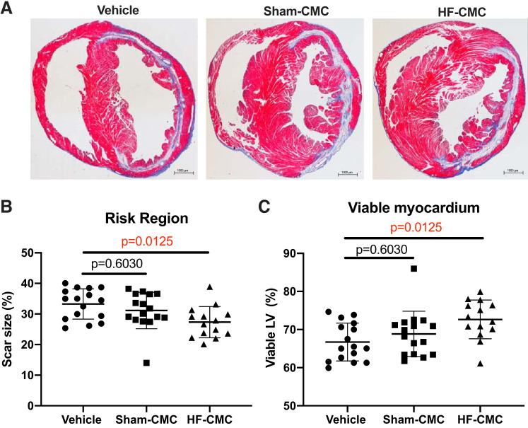
Fig. 4.
Heart failure-derived cardiac mesenchymal cells (HF-CMC) significantly reduced scar size and limited loss of viable myocardium. Sections from saline (vehicle; n = 16)-, sham (Sham-CMC; n = 16)-, and heart failure (HF-CMC; n = 14)-treated hearts were stained with Masson’s trichrome to determine scar size. A: representative images of vehicle, Sham-CMC, and HF-CMC treated hearts. B: quantification of scar size in vehicle-, Sham-CMC-, and HF-CMC-treated hearts. C: quantification of viable myocardium in vehicle, Sham-CMC, and HF-CMC treated hearts. Data are represented as means ± SD. One-way ANOVA followed by Sidak’s multiple-comparison test was used to determine significant differences between Sham-CMC- or HF-CMC-treated hearts compared with vehicle.

