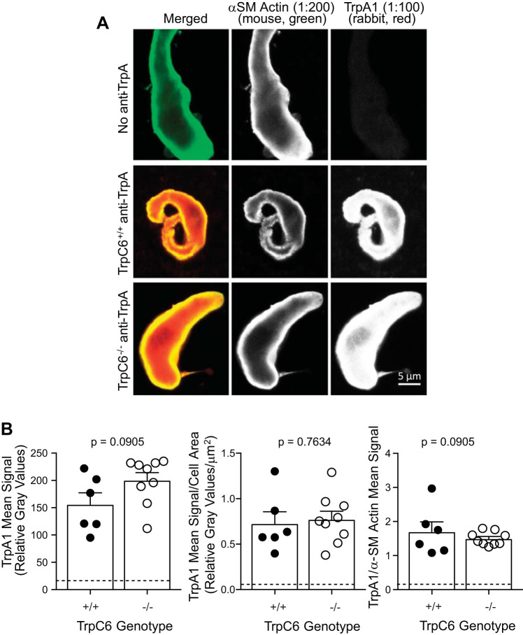
Fig. 6.
TrpA1 expression is not altered in cerebral vessels and vascular smooth muscle cells (VSMCs) from TrpC6−/− mice. Representative (A) and group (B) data for TrpA1 immunolabeling in isolated cerebral VSMCs. A: immunolabeling for TrpA1 in representative dissociated cerebral VSMCs is shown. Single-color images are shown in white to aid in visualization of intensity gradations. The first column represents α-smooth muscle (SM) actin labeling in (green in merged image), the middle column represents TrpA1 labeling (red in merged image), and the last column represents a merged image of α-SM and TrpA1. Top row: dissociated cerebral VSMC from a TrpC6−/− animal labeled with α-SM actin and both secondary antibodies, but no rabbit anti-TrpA1 antibody, served as a negative control. Middle row: VSMC from a TrpC6+/+ animal showing TrpA1 expression is robust and associated with α-SM actin which is localized just below the membrane in freshly isolated VSMCs. Bottom row: VSMC from a TrpC6−/− animal showing similar localization and levels of TrpA1. B: quantitation of TrpA1 signaling alone and normalized to cell area and α-SM actin are shown. No differences in TrpA1 signal were found. Data were analyzed using a 2-tailed independent t test, P values are provided.

