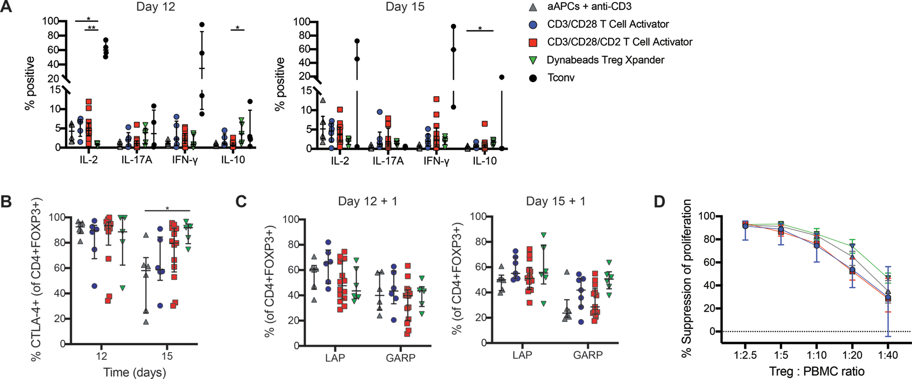
Figure 3: Function of thymic Tregs expanded with cell-free activation reagents.

Isolated thymic Tregs were expanded with the indicated type of activation reagent and analyzed by flow cytometry after 12 or 15 days. Expression of (A) intracellular cytokines in Treg (CD4+FOXP3+) and Tconv after 4 hours of activation with PMA, ionomycin and brefeldin A, (B) CTLA-4 or (C) LAP and GARP after 24 hours of activation with anti-CD3/CD28 beads at a 1:16 bead to cell ratio. (D) After 15 days of expansion, thymic Tregs were cocultured with cell proliferation dye (CPD)-labeled PBMC at the indicated ratios and stimulated with a 1:16 ratio of anti-CD3/CD28 beads for 4 days. Suppression of CD8+ T cells within PBMC was determined by division index. For A-C, within each group, each symbol represents cells from a different subject and bars indicate median ± interquartile range. For D, median ± interquartile range is shown. n=4–6 for L cell aAPCs + anti-CD3 mAbs, n=4–6 for CD3/CD28 T Cell Activator, n=4–14 for CD3/CD28/CD2 T Cell Activator, n=2–6 for Dynabeads Treg Xpander, and n=3–4 for Tconv all tested in 4–9 individual experiments. *P < 0.05, **P < 0.01 as determined by a (A) Kruskal-Wallis test with Dunn’s multiple comparisons test to compare expression for each cytokine among activation reagents or (B) two-way ANOVA with Tukey’s multiple comparisons test to compare CTLA-4 expression between conditions on each day.
