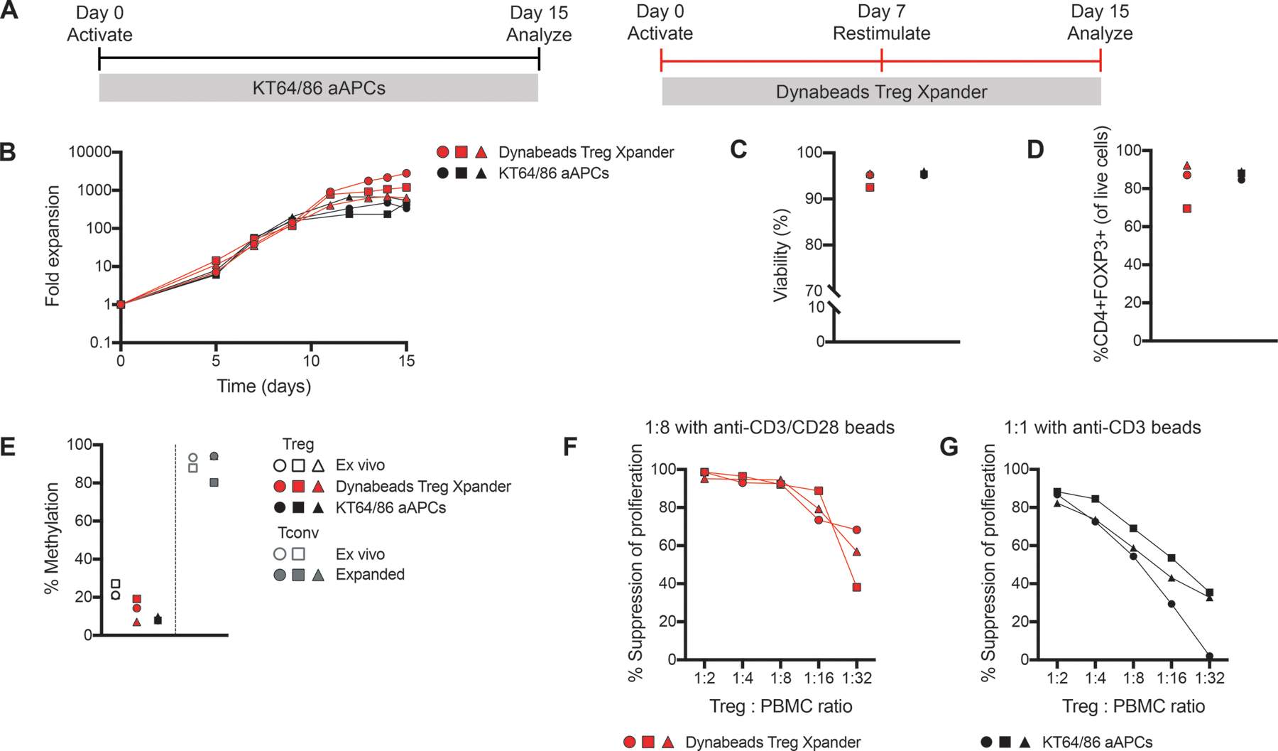
Figure 5: Thymic Treg expansion with KT64/86 aAPCs.

Replicate aliquots of thymic Tregs were expanded using KT64/86 aAPCs or Treg Xpander as indicated in (A). (B) Cells were counted to determine fold expansion. After 15 days, cells were analyzed for (C) viability and (D) FOXP3 expression. (E) TSDR analysis of ex vivo and expanded thymic Tregs and Tconv; all data are from males and the average methylation for 7 CpGs within the TSDR is shown. (F-G) Expanded thymic Tregs were cocultured with CPD-labeled (F) or CFSE-labeled (G) PBMC at the indicated ratios and stimulated at 1:8 with anti-CD3/CD28 beads or (F) 1:1 with anti-CD3 beads (G) for 4 days. Suppression of CD8+ T cells within PBMC was determined by division index. Within each group, each symbol represents cells from a different subject and matched subjects are shown with the same symbol. n=3 from 1–3 experiments.
