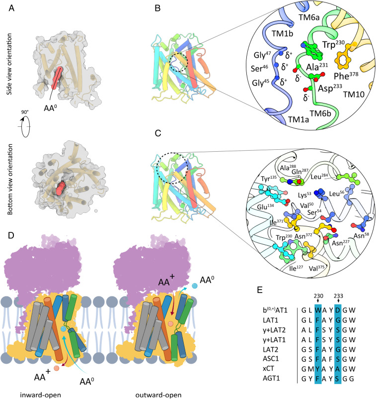
Fig. 3.
Substrate-binding site and extracellular barrier of b(0,+)AT1. (A) Surface representation of b(0,+)AT1 in side and top view orientations. The red volume indicates a substrate-accessible channel connecting the cytoplasm and the AA0-binding pocket. (B) Tube model depiction of the inward-open b(0,+)AT1 conformation and close-up view of the putative AA0-binding pocket formed by TMs 1 (blue), 6 (green), and 10 (yellow). (C) General location and close-up view of the extracellular barrier formed by TMs 1b (blue), 3 (turquoise), and EL4 (green). (D) Schematic presentation of transmembrane segments involved in cytoplasmic and extracellular barrier formation during a AA+/AA0 transport cycle. (E) Sequence comparison of residues involved in formation of substrate-binding sites between selected members of HAT-associated human SLC7 transporters. AA+: cationic amino acid. AA0: neutral amino acid.

