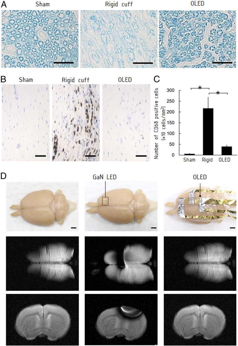
Fig. 3.
Characterization of OLED attached on neuronal tissues. (A) Histological sections of sciatic nerves with a cuff representing a rigid optical emitter and the OLED attached around the nerve for 10 d (Materials and Methods). The transverse section stained with Luxol fast blue exhibits significant morphological change of the myelin sheath when the rigid cuff was attached. There was no clear difference between the nerve with the OLED and the sham-operated nerve. (Scale bars, 20 μm.) (B) CD68 immunostaining on the longitudinal sections. The nerve with the rigid cuff exhibited overexpressed CD68, suggesting damage to the nerve. The nerve with the OLED did not exhibit overexpression. (Scale bar, 50 μm.) (C) The number of CD68-positive cells per unit area (cells per square millimeter) in each group (sham, rigid cuff implantation, and OLED implantation). Data are presented as means ± SEM. Asterisk denotes statistically significant differences across the groups (one-way ANOVA, F = 7.32, P = 0.02; Tukey–Kramer HSD test, P < 0.05). (D) MRI of a perfusion-fixed rat brain and the brain with attached conventional GaN LED and OLED. (Scale bar, 2 mm.) While the GaN LED caused an artifact, the influence of the OLED was negligible.

