Figure 2.
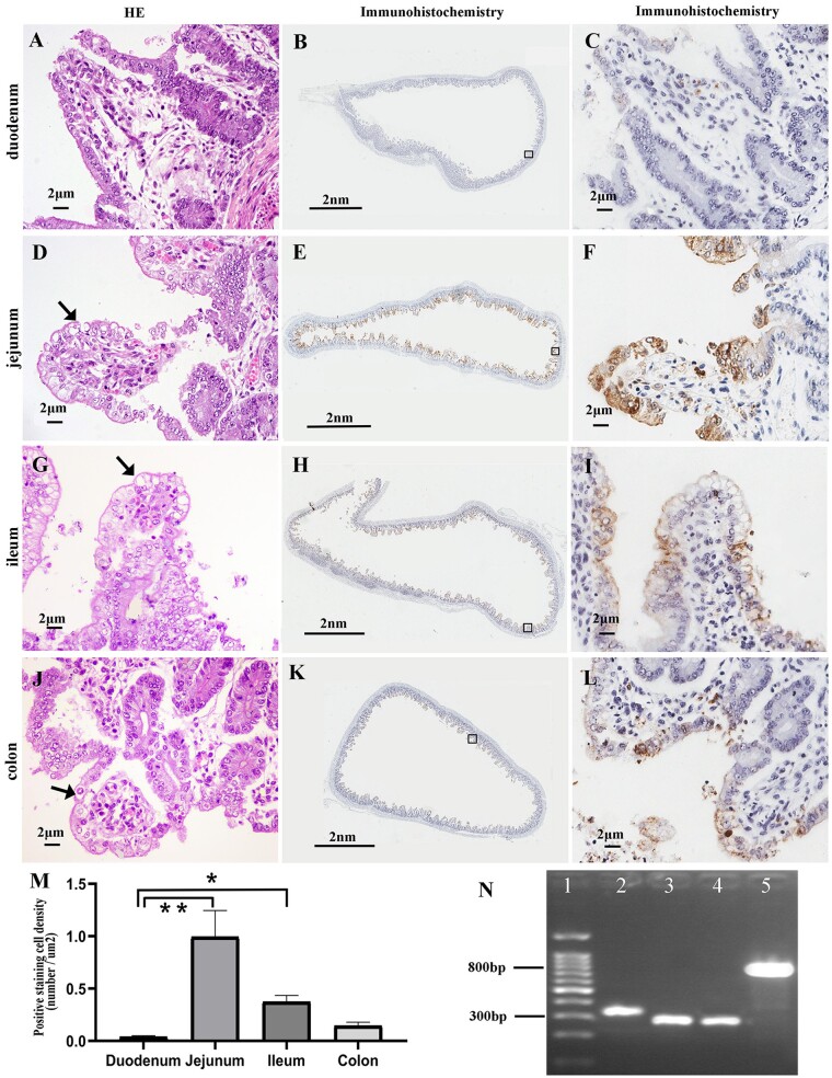
Histopathological features, PEDV antigen distribution, and PCR results. (A), (D), (G), and (J) Intestinal histopathological features of the affected piglets in H & E stained sections. Vacuolated epithelial cells are indicated by arrows. (B), (C), (E), (F), (H), (I), (K), and (L) show IHC staining to detect PEDV antigen in the different intestinal tracts of one infected piglet. The approximate locations of the first and third column tissues are indicated by the black squares in the second column tissue. (M) IHC analysis showing the staining density in different tissues, **P < 0.01, *P < 0.05. (N) PCR results: lane 1, marker; lane 2, field strain positive control; lane 3, vaccine strain positive control; lane 4, ORF3 gene detection of sample strain; lane 5, S gene detection.

