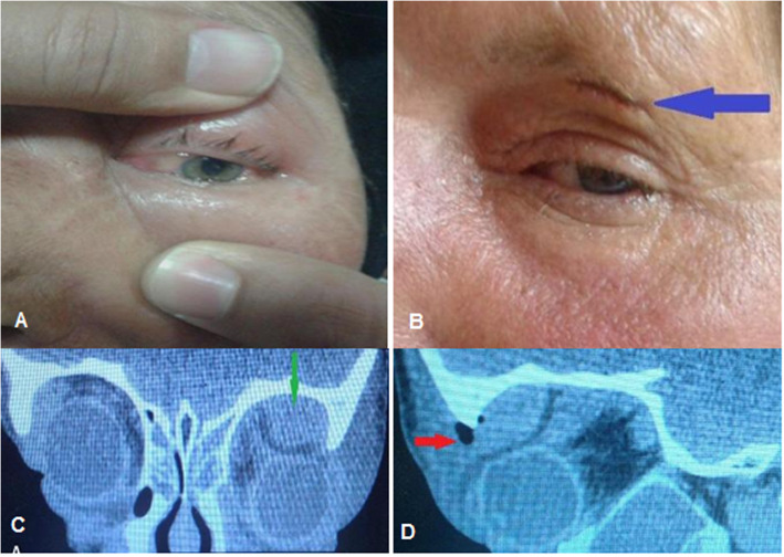
Fig. 7.
A 62-year-old woman with visual impairment in presenting time, a external eye exam shows upper lid swelling and redness with limitation of motion and conjuctival chemosis, b 5 day after external abscess drainage with sub-brow incision (blue arrow), c orbital CT scan coronal view revealed a left supra-temporal subperiostal abscess (green arrow), d and sagittal view showed a superior abscess with hypodense area (red arrow) and compression effect to the globe

