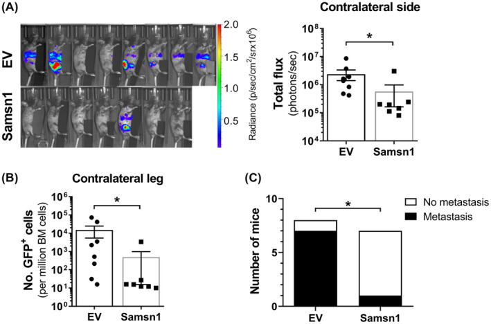
FIGURE 2.

Samsn1 inhibits the metastasis of 5TGM1 cells in vivo. 5TGM1‐Samsn1 or 5TGM1‐EV cells were injected into the left tibia of KaLwRij mice and tumor burden was measured by BLI and flow cytometry. (A) BLI scans of the contralateral side (injected leg covered) of the mice inoculated with 5TGM1‐EV or 5TGM1‐Samsn1 cells and the quantitated total fluxes after 23 days are shown. (B) The number of GFP+ tumor cells in the BM from the non‐injected, contralateral leg was assessed by flow cytometry after 23 days. (C) The number of mice injected i.t. with 5TGM1‐EV or 5TGM1‐Samsn1 cells with overt metastasis, defined as visible BLI signal from sites other than the injected leg and/or greater than 200 tumor cells per million in the BM of the contralateral leg by flow cytometry. Results were normalized to primary tumor burden and graphs depict the mean ± SEM of n = 7‐8 mice per cell line from two independent experiments. *p < 0.05, Mann–Whitney U test (A and B) or Fisher's exact test (C)
